
Introduction
The hip dysplasia is one of the most common hereditary orthopedic disorders in dogs. It mainly affects large and giant breeds, as well as some medium-sized breeds. Hip dysplasia is a developmental pathology of inherited origin. multifactorial and polygenic. It is characterized by joint laxity at the hip, leading to structural abnormalities such as subluxations and microfractures, resulting in osteoarthritis over time. Early detection and appropriate management are essential to limit disease progression and improve the comfort of canine patients.
Aetiology and predisposed breeds
Hip dysplasia is a genetically transmitted genetically transmitted which mainly affects large dogs, although some breeds of cat, such as the Maine Cooncan also be affected (with a prevalence of 15-18%). Dog breeds most at risk include :
- Labrador Retriever
- Golden Retriever
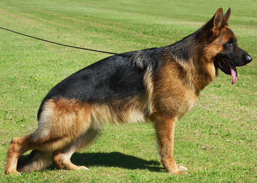
- German Shepherd
- Rottweiler
- Bulldog
- Mastiff
- Saint-Bernard
- Carlin
Visit early detection is complicated by the developmental nature of the disease. Clinical signs generally appear from the age of 4 months, although subtle signs may appear earlier.
Pathophysiology
Hip dysplasia is the result of joint joint laxity resulting in microfractures of the dorsal margin and degeneration of the femoral head and acetabulum. Pathophysiological stages include:
- Joint laxity Early detection can reveal this laxity before clinical signs are visible.
- Subluxation The head of the femur loses its alignment with the acetabulum.
- Inflammation and synovitis Inflammation of surrounding structures.
- Osteoarthritis Progressive cartilage degeneration, capsular thickening and osteophyte formation.
Anamnesis and clinical symptoms
Dogs suffering from hip dysplasia may present several clinical signs. clinical signs :
- A swaying gait and bunny hopping (jerky movement of hind legs when running).
- Intermittent lameness or permanentaggravated after exercise or when getting up.
- Amyotrophy of the thigh muscles.
- Pain on palpation and reduced joint amplitude.
Diagnostic procedures
Visit diagnosis is based on a combination of clinical and complementary examinations. The main methods include :
- Clinical examination Check for lameness, abnormal gait and reduced muscle symmetry.
- Joint laxity tests Ortolani and Barlow tests to measure joint laxity under anesthesia.
- X-rays Used to visualize subluxation and joint deformity.
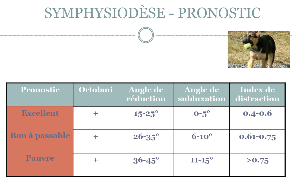
- Examination under anesthesia Allows measurement of angles of reduction (Ortolani) and subluxation (Barlow) to assess degree of dysplasia.
Processing options
Treatment options vary according to the severity of the dysplasia and the age of the animal.
Surgical treatments
- Juvenile pubic symphysiodesis (JPS) Preventive surgery in puppies aged 12 to 20 weeks to induce premature fusion of the pubic growth plate, improving acetabulum alignment.
- Triple pelvic osteotomy (TPO) Procedure performed on young dogs with moderate dysplasia, allowing ventro-lateral rotation of the acetabulum.
- Total hip replacement (THR) Therapeutic option for severe cases of osteoarthritis, with complete replacement of the joint by a prosthesis. This technique offers an excellent prognosis, with a major complication rate of less than 15%.
- Femoral head and neck osteotomy (FHO) Indicated to relieve pain in small to medium-sized dogs by removing the head and neck of the femur.
- Hip denervation Palliative technique designed to reduce pain by cutting sensory nerves in the hip.
Medical treatments
For mild cases or as a complement to surgical treatment:
- Non-steroidal anti-inflammatory drugs (NSAIDs) Reduces inflammation and relieves pain.
- Chondroprotectors (glucosamine, chondroitin): Used to improve cartilage health and delay the progression of osteoarthritis.
- Physiotherapy and rehabilitation Programs to strengthen muscles and improve joint mobility.
Prognosis and long-term management
The prognosis of hip dysplasia depends on the age of diagnosis and the severity of the disease. With early management, including surgical procedures and medical medical follow-upWith the right treatment, dogs can regain a satisfactory quality of life. Weight control and appropriate exercise also play a key role in the long-term management of the condition.
Conclusion
The hip dysplasia is a complex condition that requires early detection and multimodal multimodal management. General practice veterinarians play an essential role in detecting clinical signs and developing appropriate treatment strategies, whether preventive or curative. With advances in orthopedic surgery and physiotherapy techniques, it is possible to significantly improve the quality of life of affected dogs.

