
Introduction
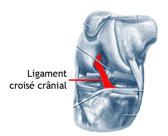
The rupture of the cranial cruciate ligament (RLCC) is one of the most common orthopedic disorders in dogs. This ligament, similar to the anterior cruciate ligament in humans, is essential for the stability of the canine knee. However, unlike in humans, where rupture is often the result of trauma, in dogs this condition is generally degenerative and chronic, although in some cases it can be acute. Rupture of the cranial cruciate ligament can lead to progressive lameness and complications such as meniscal damage and osteoarthritis. This article explores the mechanisms of the disease, diagnostic options and treatments available to general veterinarians.
Aetiology and predisposed breeds
The exact cause of cranial cruciate ligament rupture in dogs remains unknown, but is considered to be multifactorialThe result is progressive degeneration of the ligament. Here are the main contributing factors:
- Genetic predisposition Some breeds are more prone to this condition, notably Labrador Retrievers, Golden Retrievers, Bernese Mountain Dogs, Rottweilers, Boxers, Bulldogsand Mastiffs.
- Overweight Excess weight puts extra pressure on joints, increasing the risk of rupture.
- Bone conformation Certain abnormalities in bone conformation can lead to knee instability.
- Hyperactivity or poor physical condition These factors can accelerate ligament degeneration.
Clinical symptoms
The clinical signs of cranial cruciate ligament rupture can vary according to the severity of the injury. Initial symptoms are often subtle, but may develop over time if knee instability worsens. The most common symptoms include:
- Intermittent lameness or persistent lameness after physical activity.
- Difficulty getting up or getting into a car.
- Abnormal posture sitting with hind leg extended to the side.
- Reduced physical activity and muscle loss in the affected thigh.
- Pain when manipulating the knee.
- Clunking” noise on walkingoften associated with meniscus damage.
- Swelling of the knee and progressive development of osteoarthritis, leading to stiffness and persistent lameness.
In more advanced cases, complete rupture of the cranial cruciate ligament causes severe lameness and, often, non-weight-bearing on the affected limb.
Diagnosis of Cranial Cruciate Ligament Rupture
Diagnosis is based on several clinical and radiographic steps. Methods used include :
- Clinical examination and palpation Palpation of the knee can detect joint instability. The anterior drawer and the tibial compression test are key techniques for assessing complete ligament rupture.
- X-rays They can be used to assess signs of joint inflammation (synovial effusion) and to visualize osteoarthritis. They are also useful for ruling out other pathologies such as bone tumours.
- MRI or arthroscopy These techniques allow direct visualization of the cruciate ligament and menisci, and are often used to plan surgery.
Processing options
Treatment of cranial cruciate ligament rupture depends on the severity of the rupture, the dog’s age, weight and activity level. Options include surgical and non-surgical treatments.
Surgical treatment
Surgery is recommended for the majority of dogs, especially those of medium and large size, to stabilize the joint and prevent the progression of osteoarthritis. The main surgical techniques include:
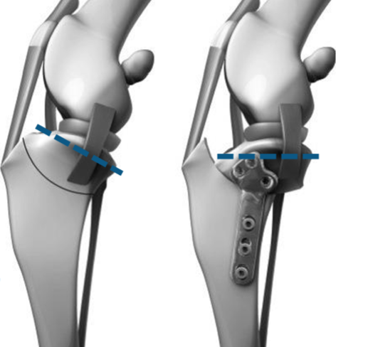
- Tibial Plateau Leveling Osteotomy (TPLO) : This is the most effective long-term surgical technique. It modifies the biomechanics of the knee by levelling the tibial plateau, thus providing greater stability without replacing the cranial cruciate ligament.
- Fabellar lateral suture This extra-articular technique is used to stabilize the joint in small to medium-sized dogs. It is generally reserved for dogs weighing less than 20-30 kg.
- Tightrope A recent technique that replaces the cruciate ligament with a synthetic suture to stabilize the joint.
Non-surgical treatment
In some dogs (small breeds, inactive dogs or those with surgical contraindications), conservative treatments may be considered:
- Activity restriction and anti-inflammatories Limiting the dog’s activity while administering anti-inflammatory medication can temporarily relieve lameness. However, osteoarthritis often progresses despite these measures, and persistent lameness is likely.
- Physiotherapy Post-surgical or non-surgical rehabilitation programs can improve the animal’s recovery and quality of life.
Prognosis and post-operative management
The prognosis for dogs undergoing TPLO surgery is generally good, with a return to normal activity in 85-90% of cases. Large or very active dogs benefit greatly from this procedure. The post-operative management includes :
- Strict rest for 6 to 8 weeks after surgery.
- Rehabilitation and physiotherapy exercises to speed up recovery.
- Anti-inflammatory and analgesics to manage post-surgical pain and inflammation.
Conclusion
The rupture of the cranial cruciate ligament ligament is a complex condition that requires early diagnosis and surgical surgical management adapted to offer the animal optimum quality of life. General veterinarians should be alert to subtle clinical signs in predisposed breeds, and consider early referral for surgery, particularly in larger dogs. With modern surgical techniques such as TPLO, the long-term prognosis is favorable.

